

Neurosurgery and Neurology · Jaipur Hospital
Laminectomy in Jaipur
Leg pain and numbness limiting your walk? We assess your spine and give a clear plan.
- Serving Jaipur since 1991 (35 years)
- NABH accredited
- 24×7 emergency & ICU
- Cashless: RGHS, MAA Yojana, CGHS + 22 insurers
- Operation time
- 2 to 3 hours
- Anaesthesia
- General anaesthesia
- Recovery
- Back to desk work in 4 to 6 weeks
- Price
- ₹1,50,000 to ₹3,00,000typical range in India; ask us for an estimate
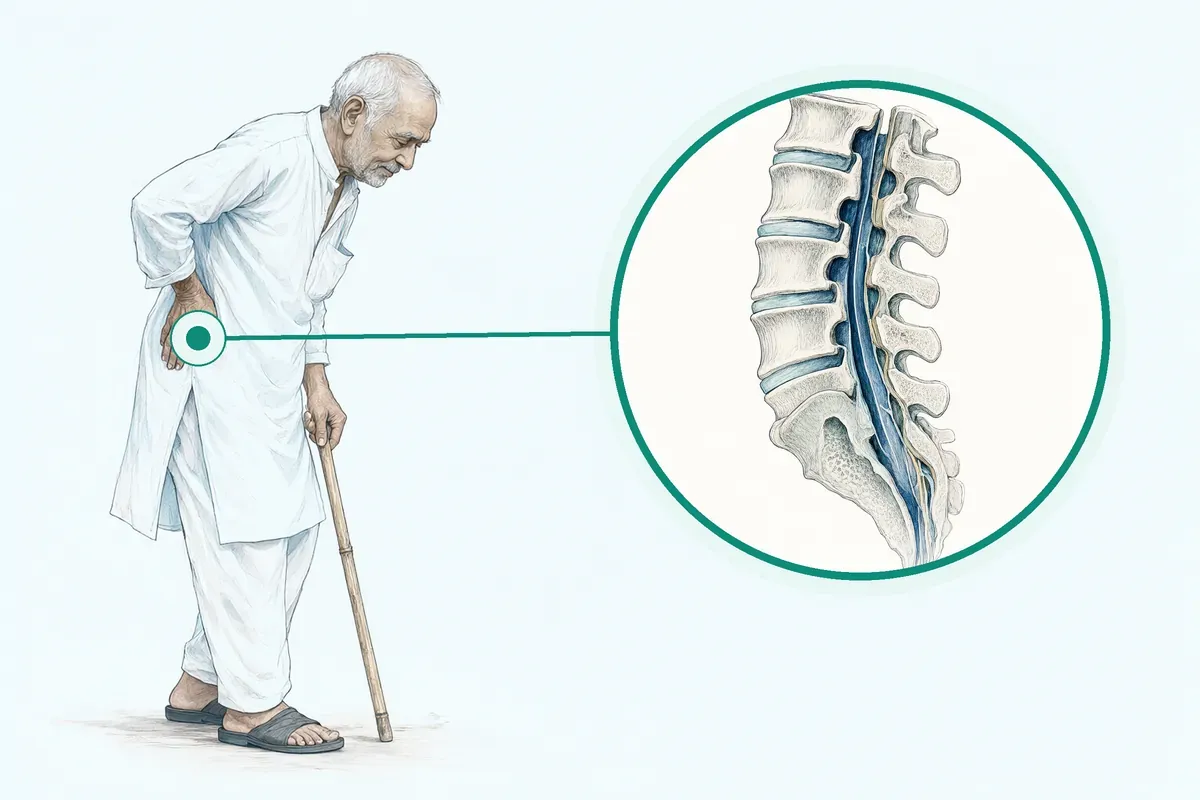
Narrowed spinal canal presses nerves
Your spinal canal has narrowed and crowds the nerves
The space inside your lower spine shrinks over time. Bone and tissue press on nerves, causing pain, numbness or heaviness in the legs when you stand or walk.
-
Leg pain on walking
Pain, tingling or weakness in the legs appears after a short distance and eases when you sit or bend forward.
-
Daily tasks become hard
Standing in the kitchen, walking to the market or climbing stairs becomes difficult and tiring.
-
Medicines and therapy not helping
Painkillers, ointments and exercises have been tried for weeks or months without lasting relief.
-
Sleep and mood affected
Night pain disturbs rest. You feel frustrated because you cannot do the things you used to do.
Lumbar spinal stenosis is the gradual narrowing of the spinal canal that crowds nerve roots and the spinal cord.
Why Jaipur Hospital
You worry that the leg pain will keep you from earning, caring for your family, or simply walking to the temple without having to stop and sit.
Your care plan
Your first steps with us
- 1 Call or visit OPD for a neurosurgeon consultation
- 2 Get scans and tests done in-house the same day
- 3 Review options with your surgeon and receive a cost estimate
- 4 Have surgery, recover in hospital, then continue physiotherapy at home
How the operation is done
Under general anaesthesia, the surgeon removes the bony arch (lamina) to widen the spinal canal and relieve pressure on the nerves. The procedure usually takes two to three hours.
-
Open Laminectomy
Standard posterior approach with full lamina removal to decompress the spinal canal.
-
Bilateral Laminectomy
Both sides of the lamina are removed for central stenosis affecting both nerve roots.
-
Laminectomy with Fusion
Pedicle screws are added if the spine is unstable or a spondylolisthesis is present.
Why patients trust us
- 1991 Serving Jaipur for 35 years
- NABH Accredited hospital
- 1 Neurosurgery and Neurology consultants
- 24×7 Emergency, ICU and ambulance
- 25 Cashless schemes and insurers
What research shows about decompressive laminectomy
-
7.8 points better ODI score at 2 years
In a randomised trial, surgery improved disability scores more than non-operative care at two years.
-
No difference in ODI at 2 and 5 years with or without fusion
Adding fusion to decompression did not give better disability outcomes at two or five years.
-
Complication rate 10% to 24% in surgical groups
Studies reported complications such as haematoma, infection, or reoperation in 10 to 24 out of 100 surgical patients.
If it is left untreated
Without treatment, about one-third of patients improve on their own, half stay the same, and 10 to 20 percent get worse over three years.
Life after treatment
Most people return to desk work in four to six weeks and walk farther with less leg pain.
-
Walk longer distances
Most people find they can walk further before leg pain or heaviness forces them to stop.
-
Return to daily routine
Usual activities like marketing, cooking and light housework become manageable again after recovery.
-
Less need for pain medicines
Many patients reduce or stop daily painkillers once the nerve pressure is relieved.
-
Better sleep and mood
With night pain reduced, sleep improves and you feel more like yourself again.

Frequently asked questions
Questions patients ask about laminectomy
What does lumbar canal stenosis decompression involve?
In short
Lumbar canal stenosis decompression at Jaipur Hospital involves removing the lamina to relieve pressure on the spinal cord and nerves. The surgeon makes an incision in the lower back, removes the bony arch and any bone spurs, and widens the spinal canal. The operation usually takes two to three hours under general anaesthesia.
In detail
A laminectomy is surgery on your spine to remove the lamina, the bony arch that protects the back part of your spinal cord. Removing the lamina relieves pressure on your nerves and spinal cord by making more room in the spinal canal.
The surgeon may also remove bone spurs that strain the nerve roots. This procedure treats conditions like spinal stenosis and herniated disks by decompressing the affected nerves. The type of procedure used depends on the diagnosis and severity.
Open laminectomy uses a standard posterior approach with full lamina removal, while bilateral laminectomy decompresses both sides for central stenosis. If instability is present, fusion with pedicle screws may be added.
A common belief
Many people think the surgeon always fuses the spine with screws.
What actually happens
Fusion is only added if there is proven instability, such as spondylolisthesis. Studies show decompression alone gives similar long-term outcomes to decompression with fusion for many patients, while fusion carries higher risks of blood loss, infection, and longer hospital stays.
Your next step at Jaipur Hospital
You will meet your surgeon in the OPD for a clinical assessment and imaging review. Pre-operative tests include blood work, ECG, and MRI or CT scans. Hospital stay is typically three to five nights. You can usually return to desk work in four to six weeks.
Which tests and scans do I need before lumbar canal stenosis decompression?
In short
Before lumbar canal stenosis decompression at Jaipur Hospital, you will need an MRI of the lumbar spine to confirm the level and severity of canal narrowing. The surgeon also orders routine pre-operative tests: blood counts, kidney and liver function, coagulation profile, blood sugar, ECG, and chest X-ray to assess fitness for general anaesthesia.
In detail
Diagnosis of lumbar spinal stenosis is confirmed with cross-sectional imaging, typically MRI, which shows the narrowed spinal canal and compressed nerves. Clinical history of neurogenic claudication — leg pain on walking relieved by sitting — must match the imaging findings.
Pre-operative blood work and cardiac screening are standard for any patient undergoing general anaesthesia. If you have other medical conditions, additional clearance from a physician or cardiologist may be required. The surgeon will also review any medications you take, especially blood thinners, which may need to be stopped temporarily.
A common belief
X-rays alone cannot show nerve compression.
What actually happens
MRI is the standard. Some patients think a CT scan replaces MRI, but MRI gives better soft tissue detail for surgical planning.
Your next step at Jaipur Hospital
Bring all prior imaging and medication lists to your pre-operative appointment. Plan for a three to five night hospital stay. Recovery to desk work takes four to six weeks. The typical cost range is ₹1,50,000 to ₹3,00,000.
How painful is the laminectomy procedure and the early recovery?
In short
During laminectomy at Jaipur Hospital you receive general anaesthesia, so you are asleep and feel no pain during the operation. After surgery, you will have incisional pain and muscle soreness in the lower back. The nursing team gives regular pain relief through intravenous and oral medicines so most patients can move comfortably within the first day.
In detail
Evidence confirms that on the day of surgery an anaesthesiologist gives you general anaesthesia and you are asleep for the entire procedure without feeling pain. Post-operative pain is managed with a multimodal approach including opioids, anti-inflammatory drugs, and sometimes nerve blocks.
Studies comparing surgical techniques show that open decompression causes more early pain and higher opioid use than minimally invasive approaches, but both methods achieve good pain control with standard protocols. Early mobilisation is encouraged because it reduces stiffness and speeds recovery.
Your pain score is checked frequently and medication is adjusted so you can sit, stand, and walk short distances as soon as possible.
A common belief
Some patients expect either no pain at all or unbearable pain.
What actually happens
The reality is moderate, manageable pain that improves steadily. Staying still because of fear of pain actually slows recovery.
Your next step at Jaipur Hospital
Expect to stay in hospital for three to five nights. Pain medicine continues at discharge with a tapering schedule over two to three weeks.
You will receive instructions on wound care, activity limits, and when to contact the hospital if pain worsens or new symptoms appear. Most people return to desk work in four to six weeks.
How long does recovery usually take after laminectomy?
In short
After laminectomy at Jaipur Hospital, most people return to desk work in four to six weeks. The hospital stay is typically three to five nights. Light walking starts the day after surgery. Heavy lifting, bending, and twisting are restricted for six to eight weeks. Full return to strenuous activity or manual labour may take three months or more, depending on your job and healing.
In detail
Evidence indicates that operation time is two to three hours and hospital stay averages three to five nights.
Studies comparing surgical techniques show that minimally invasive approaches have shorter mobilisation times and faster early recovery than open decompression, but both achieve similar functional outcomes at one to two years.
In a non-operative series, about one-third of patients improved over three years, half stayed the same, and 10% to 20% worsened. Surgery generally provides better leg pain and disability outcomes than continued non-operative care. Your surgeon will guide a stepwise increase in activity based on wound healing, pain control, and muscle strength.
A common belief
Some expect to be fully active in two weeks, others fear months of bed rest.
What actually happens
The truth is a gradual return: walking early, lifting later, with steady improvement guided by your surgeon.
Your next step at Jaipur Hospital
Plan for an attendant to help at home for the first one to two weeks. Follow the physiotherapy programme given at discharge. Attend scheduled follow-up visits for wound check and progress review. Most patients resume driving when off strong pain medicines and can turn comfortably, usually around three to four weeks.
Does the clinical examination match the findings on my scan for lumbar canal stenosis?
In short
Your surgeon compares your symptoms and physical signs with the MRI or CT images to decide if surgery is appropriate. Jaipur Hospital follows the standard practice of correlating clinical findings such as walking distance, reflexes and sensory changes with the degree of canal narrowing seen on imaging. A scan alone does not determine the need for laminectomy; the decision rests on how much the stenosis affects your daily life.
In detail
Evidence shows that many people have radiological stenosis without symptoms, while others have severe symptoms with only moderate narrowing on scans.
The diagnosis of lumbar spinal stenosis is made when the clinical history of neurogenic claudication — pain brought on by standing or walking and relieved by sitting or bending forward — matches the imaging.
Your surgeon tests muscle strength, sensation, reflexes and straight-leg raise to map the affected nerve roots. Only when the examination and scan point to the same level and side is decompressive laminectomy considered. This careful matching avoids unnecessary surgery on incidental findings.
A common belief
A common belief is that a bad-looking scan means you need surgery.
What actually happens
In reality, many people have stenosis on imaging but no trouble walking. Operating on the scan alone can lead to unnecessary procedures. The clinical examination is what links the image to your actual problem.
Your next step at Jaipur Hospital
Bring all your imaging reports and films to the consultation. The surgeon will review them with you and explain the correlation. If there is a mismatch, further tests such as nerve conduction studies or a different scan may be ordered.
The final plan is made together with you. To have your scans and symptoms assessed by the surgical team, book a consultation at Jaipur Hospital.
What changes in strength, sensation, speech, vision, or bladder control need urgent assessment after laminectomy?
In short
After laminectomy, seek urgent care for new or worsening leg weakness, foot drop, numbness in the saddle area (inner thighs, buttocks, genital region), inability to pass urine or control bowels, or loss of sensation when wiping. Jaipur Hospital treats these as possible nerve injury, haematoma or cauda equina syndrome requiring immediate imaging and possible re-operation. Speech and vision changes are not typical of lumbar surgery but should still be reported at once.
In detail
The surgical site is near the cauda equina nerve roots. A post-operative haematoma or epidural abscess can compress these roots within hours to days, causing saddle anaesthesia, urinary retention, incontinence and bilateral leg weakness.
Evidence reports complication rates of 10 to 24 percent in surgical series, including haematoma and nerve injury. Early detection and decompression improve recovery chances. Normal post-operative symptoms include incisional pain, some leg aching from nerve root manipulation, and temporary numbness around the wound.
Any new neurological deficit that was not present before surgery or that worsens after initial improvement is a red flag. Speech or vision changes may indicate an unrelated neurological event and also need emergency evaluation.
A common belief
Patients often think all post-surgery pain or numbness is normal.
What actually happens
Incisional pain and some leg aching are expected. But new weakness, saddle numbness or bladder changes are not normal — they are emergencies. Knowing the difference can save nerve function.
Your next step at Jaipur Hospital
Keep the emergency numbers handy: 0141-2551500, 0141-2554139, +91-7414-001-001. If you notice any red-flag symptom, call or come to the 24×7 emergency department immediately. Do not wait for the morning OPD. Your attendant should be aware of these signs. For clarification on what is normal versus urgent during your recovery, book a consultation at Jaipur Hospital.
Why might my doctor recommend lumbar canal stenosis decompression?
In short
Your doctor at Jaipur Hospital may recommend lumbar canal stenosis decompression if you have persistent leg pain, numbness, or walking difficulty caused by narrowing of the spinal canal. Surgery is considered when symptoms limit daily activities and do not improve with non-surgical treatments like medicines and physiotherapy.
In detail
Lumbar spinal stenosis is a gradual narrowing of the spinal canal that crowds nerve roots and the spinal cord. This may cause severe pain, numbness, tingling, or weakness in the legs, and limit the distance you can walk.
The condition often worsens with standing or walking and improves with sitting or forward bending. First-line therapy is activity modification, analgesia, and physical therapy. Most patients are treated non-operatively.
However, surgery appears effective in carefully selected patients with back, buttock, and lower extremity pain who do not improve with conservative management. A laminectomy may be an option to help relieve or reduce these symptoms when they are sufficiently bothersome and persistent.
A common belief
A common belief is that surgery is needed as soon as stenosis is seen on an MRI.
What actually happens
In reality, many people have stenosis on scans but few symptoms. Studies show symptoms often wax and wane, and rapid deterioration is rare. Surgery is almost always elective, not an emergency, and is offered only after a fair trial of non-surgical care.
Your next step at Jaipur Hospital
The surgeon will review your MRI or CT scan alongside your symptom history. If non-surgical measures have failed over several weeks or months, a surgical opinion is appropriate. You can discuss the typical hospital stay of three to five nights and recovery timeline.
How should I prepare in the days before lumbar canal stenosis decompression?
In short
In the days before lumbar canal stenosis decompression at Jaipur Hospital, follow your surgeon's specific instructions. Inform the team about all medicines and supplements you take. You may be asked to stop blood-thinning medicines several days before surgery. Do not stop any medicine unless your doctor approves. If you use tobacco, try to stop, as nicotine impairs healing.
In detail
Your surgeon will give you specific preparation instructions. You should disclose all medications, supplements, and tobacco use. Certain medicines like blood thinners may need to be stopped or adjusted to reduce bleeding risk.
Nicotine from tobacco products changes how your body heals and increases complication risk. You will attend a pre-anaesthesia check-up where the anaesthetist reviews your health and explains the anaesthesia plan. Arrange for an attendant to stay with you during your hospital stay of three to five nights.
A common belief
A common mistake is stopping medicines on your own.
What actually happens
Never stop a prescribed medicine unless your doctor tells you to. Another oversight is not planning for help at home after discharge. You will need assistance with daily tasks for the first couple of weeks.
Your next step at Jaipur Hospital
Complete all pre-operative tests as scheduled. Follow fasting instructions given by the anaesthesia team. Pack comfortable clothing, your regular medicines, and any walking aids you use. Plan for help at home after discharge, especially for the first two weeks.
For detailed preparation guidance tailored to your health, book a consultation with our neurosurgery or orthopaedics team at Jaipur Hospital.
What are the common side effects and the less common serious complications of laminectomy?
In short
Common side effects after laminectomy at Jaipur Hospital include incisional pain, back muscle stiffness, temporary numbness near the wound, and fatigue for a few weeks. Less common but serious complications can include infection, bleeding or haematoma, dural tear with cerebrospinal fluid leak, nerve injury, blood clots in the legs or lungs, and risks related to general anaesthesia.
In detail
Evidence from surgical studies reports complication rates between 10% and 24% in surgical groups. Specific complications documented include spinous process fracture, coronary ischaemia, respiratory distress, haematoma, stroke, reoperation risk, and death due to pulmonary oedema.
Fusion procedures carry higher risks of blood loss, infection, longer hospital stays, and higher costs compared to decompression alone. One study noted a participant needing revision surgery nine months after the initial operation.
The surgical team takes multiple precautions: antibiotics before incision, careful haemostasis, nerve monitoring when indicated, and early mobilisation to prevent clots.
A common belief
Many people focus only on paralysis risk, which is rare.
What actually happens
More frequent issues like wound infection or blood clots are often overlooked. Fusion adds more risk than decompression alone.
Your next step at Jaipur Hospital
Before surgery, your surgeon will explain the risks that apply to your specific case. After discharge, watch for increasing wound redness, swelling, fluid leakage, fever, new leg weakness, numbness, or difficulty passing urine. These need urgent evaluation. Routine follow-up visits are scheduled to check healing.
Which symptoms are expected after laminectomy, and which need urgent medical attention?
In short
Expected symptoms after laminectomy at Jaipur Hospital include incisional pain, back stiffness, mild swelling around the wound, temporary numbness near the scar, and fatigue for a few weeks. Leg pain often improves quickly but may fluctuate. Urgent symptoms are fever above 38°C, increasing wound redness or discharge, new or worsening leg weakness, new numbness in the groin or inner thighs, loss of bladder or bowel control, severe headache, calf swelling or pain, and shortness of breath.
In detail
Evidence shows that surgical complications such as haematoma, dural tear, infection, and nerve injury occur in a minority of patients. Studies report complication rates of 10% to 24%, including serious events like stroke and pulmonary oedema.
A dural tear can cause cerebrospinal fluid leak, presenting as a persistent clear wound discharge or positional headache. Nerve injury may cause new weakness or numbness. Blood clots can cause calf pain or sudden breathlessness.
Cauda equina symptoms — saddle numbness and bladder or bowel changes — need immediate surgery. The team monitors you closely in hospital and gives clear written instructions for home.
A common belief
Patients often dismiss new leg weakness as normal soreness or wait days with a leaking wound.
What actually happens
Both can signal serious complications that need immediate treatment.
Your next step at Jaipur Hospital
Keep the emergency numbers from Jaipur Hospital handy. If any urgent symptom appears, go to the nearest emergency department or call the hospital’s 24×7 line. Do not wait for the next scheduled follow-up. Routine follow-up is usually at two weeks for wound check and at six weeks for functional assessment.
FAQ library
Questions and answers about laminectomy
When you contact us
Get your spine assessed this week
Contact Jaipur Hospital for a neurosurgeon consultation, in-house scans, a clear explanation of options and a cost estimate before you decide.
- A consultation with a neurosurgeon in the OPD
- Your scans and reports reviewed, with tests done in-house if you need them (laboratory, X-ray and imaging)
- Your options explained in plain words, including whether you need surgery at all
- A cost estimate before you decide
- Help with cashless treatment under RGHS, MAA Yojana, CGHS and empanelled insurers and TPAs
- Follow-up visits after your treatment
Overview
Laminectomy: what to know
What lumbar spinal stenosis is and how it happens
- Discs lose height
- Discs dry out and shrink with age
- Joints and ligaments thicken
- Arthritis enlarges facet joints; ligaments stiffen
- Bone spurs form
- Extra bone grows into the spinal canal
- Herniated disc
- Soft disc material pushes through the outer covering
Lumbar spinal stenosis is a narrowing of the spinal canal in your lower back. The spinal canal is the tunnel that holds your spinal cord and nerve roots. The lamina is the bony arch that forms the back wall of this tunnel. Over time, the space inside the tunnel can become smaller. This narrowing crowds the nerves and the spinal cord.
The condition often develops as your body ages. Tissues in the spine slowly wear down. The discs between your vertebrae lose water and height. The joints and ligaments can thicken. Bone spurs may form on the vertebrae. All of these changes reduce the room available for the nerves.
A herniated disc can also narrow the space. This happens when the soft inner part of a disc pushes through its outer covering. The disc material can then press against a spinal nerve or the spinal cord.
When the nerves are compressed, they cannot work normally. This leads to pain, numbness, or weakness that may travel from your back into your buttocks and legs.
Symptoms of lumbar spinal stenosis and warning signs that need urgent care the same day
- Loss of bladder or bowel control
- Numbness in inner thighs and groin
- Sudden severe weakness in both legs
The most common symptom is pain in the buttocks or lower legs, with or without back pain. The pain often gets worse when you stand or walk. It typically improves when you sit down, bend forward, or lie down.
You may feel:
- Numbness
- Tingling
- A pins-and-needles sensation in your legs or feet
Some people notice weakness in the legs. The condition may limit how far you can walk before you have to stop and rest.
Symptoms often come and go. They may gradually worsen over months or years.
Certain symptoms need medical attention on the same day. If you lose control of your bladder or bowels, or if you have numbness in the inner thighs and groin area (saddle area), go to the emergency department immediately. Sudden severe weakness in both legs is also an emergency. These can be signs of cauda equina syndrome, which requires urgent surgery to prevent lasting damage.
Causes of lumbar spinal stenosis and who is at higher risk
- Age-related wear and tear
- Discs dry out, joints enlarge, ligaments thicken, bone spurs grow
- Born with a narrow canal
- Congenital stenosis can cause earlier symptoms
- Past injury or surgery
- Scarring or instability narrows the canal later
- Arthritis and bone diseases
- Osteoarthritis, rheumatoid arthritis, Paget's disease contribute
The main cause is age-related wear and tear in the spine. As you get older, the discs dry out and shrink. The facet joints may enlarge due to arthritis. Ligaments inside the spinal canal can thicken and stiffen. Bone spurs grow into the canal. All of these reduce the space for nerves.
Some people are born with a narrower spinal canal. This is called congenital stenosis. It can make symptoms appear earlier in life.
A past spine injury or previous spine surgery can lead to scarring or instability that narrows the canal later.
Conditions such as osteoarthritis, rheumatoid arthritis, or Paget's disease of bone can also contribute.
The condition is most common in adults over 60. It is the most frequent reason for spine surgery in people over 65. Lumbar spinal stenosis affects a large number of adults worldwide.
How lumbar spinal stenosis is diagnosed: examination and tests
- History and symptom pattern Pain worse standing or walking, better sitting or bending
- Physical examination Check leg strength, reflexes, sensation, walking on heels and toes
- Match symptoms with imaging Imaging alone is not enough; many have narrowing without symptoms
Your doctor will start by asking about your symptoms and medical history. They will check how your pain changes with different positions. Pain that worsens with standing or walking and improves with sitting or bending forward is a typical pattern.
The doctor will examine your back, check your leg strength, reflexes, and sensation. They may ask you to walk on your heels and toes. This helps find which nerves are affected.
Imaging tests confirm the diagnosis. An MRI scan shows the soft tissues, discs, nerves, and the spinal canal in detail. It is the most common test used. A CT scan can show the bones clearly. Sometimes a CT myelogram is done, where dye is injected into the spinal fluid before the scan. X-rays show the alignment of the bones and can reveal instability or arthritis.
The diagnosis is based on your symptoms matching the imaging findings. Imaging alone is not enough, because many people have narrowing on scans but no symptoms.
Non-surgical treatment options for lumbar spinal stenosis and when laminectomy is advised
- about one-third Reported improvement
- about half Reported no change
- 10% to 20% Reported worsening
Most patients with lumbar spinal stenosis are treated without surgery.
First-line treatment includes:
- Activity modification
- Pain relief medicines
- Physical therapy
Activity modification means reducing activities that bring on symptoms, such as long periods of standing or walking. Using a walking stick or a frame can help you stay active while leaning forward slightly.
Medicines often used are non-steroidal anti-inflammatory drugs (NSAIDs) to reduce pain and inflammation. Your doctor may prescribe other pain medicines if needed.
Physical therapy focuses on exercises that improve flexibility, strengthen your core and back muscles, and teach you positions that relieve pressure on the nerves.
Epidural steroid injections may be offered. They can give short-term relief for some people. However, long-term benefits have not been demonstrated in studies.
There is insufficient evidence to recommend any specific type of non-surgical treatment over another.
Surgery is almost always elective. It is considered when symptoms are sufficiently bothersome and persist despite a reasonable trial of non-surgical treatments. Rapid deterioration is rare.
In studies, surgery appears effective in carefully selected patients who do not improve with conservative management. A randomised trial found that decompressive laminectomy improved symptoms more than non-operative therapy on a disability score at two years.
In a series of patients followed without surgery for up to three years, about one-third reported improvement, about half reported no change, and 10% to 20% reported worsening.
Your surgeon will discuss whether laminectomy is appropriate for you after reviewing your symptoms, examination, and imaging.
Preparing for laminectomy surgery
Do
- Complete routine tests
- List all medicines and supplements
- Stop tobacco several weeks before
- Fast from midnight before surgery
Before your laminectomy, the surgical team will ask you to complete a few routine tests. These usually include blood tests, a chest X-ray, and an ECG to check that you are fit for general anaesthesia. If you have other medical conditions, you may also see a physician or a cardiologist for clearance.
Bring a list of every medicine and supplement you take, including any blood thinners, painkillers, or herbal products. Your surgeon will tell you which ones to stop and when to stop them. Do not stop any medicine on your own unless your doctor approves it.
If you use tobacco, try to stop several weeks before the operation. Nicotine can change the way your body heals and may raise the risk of complications.
You will be asked to fast from midnight on the night before surgery. This means no food and no water after the time given by the anaesthesia team. On the morning of the operation, take only the medicines your anaesthetist has cleared with a sip of water.
Pack comfortable loose clothing, your regular toiletries, any walking aid you use, and a copy of your insurance card or scheme papers. An attendant can stay with you in the room during your hospital stay.
How laminectomy surgery is done
Laminectomy is performed under general anaesthesia. You will be asleep for the entire procedure and will not feel any pain.
On the day of surgery, you will lie face down on a special table with pads that protect the front of your body while the surgeon works on your spine.
The surgeon makes an incision in the middle of your lower back. The muscles are moved aside to reach the vertebrae. The lamina, which is the arched back piece of the vertebra, is removed to make more room in the spinal canal. This relieves pressure on the spinal cord and nerves. Bone spurs that press on the nerves are also taken out.
Your surgeon will choose the technique that fits your condition:
- Open laminectomy: the standard posterior approach with full removal of the lamina.
- Bilateral laminectomy: both sides of the spinal canal are decompressed, often used for central stenosis.
- Laminectomy with fusion: if there is instability, pedicle screws and rods are added to hold the bones together while they fuse.
The operation usually takes two to three hours. It can take longer if more than one level is treated or if fusion is added.
Sometimes laminectomy is the first part of a two-step procedure that also includes a diskectomy, foraminotomy, or spinal fusion.
Risks and possible complications of laminectomy
- 10% to 24% Complication rate range
- spinous process fracture, coronary ischaemia, respiratory distress, haematoma, stroke, need for reoperation, death due to pulmonary oedema Reported complications
Every operation carries some risk. With laminectomy, the most common risks include infection, bleeding, a leak of spinal fluid (durotomy), and a collection of blood (haematoma) near the wound. Nerve injury is possible but uncommon.
In studies that compared surgical groups, complication rates ranged from 10% to 24% of participants.
Reported complications included:
- Spinous process fracture
- Coronary ischaemia
- Respiratory distress
- Haematoma
- Stroke
- Need for reoperation
- Death due to pulmonary oedema
When fusion is added to decompression, the risk of complications such as blood loss, infection, longer hospital stays, and higher costs increases. One study reported that a participant needed revision surgery nine months after the first operation.
Your surgical team will discuss the risks that apply to your age, general health, and the exact procedure planned. They take steps during surgery and in the ward to lower these risks, such as antibiotics to prevent infection and careful monitoring of your vital signs.
Recovery and aftercare after laminectomy
After the operation you will wake up in the recovery area and then move to a ward. Nurses will check your pain level, leg movement, and wound regularly. You will be given pain relief through a drip or tablets so you can move comfortably.
Most people start walking with help on the first or second day. A physiotherapist will show you safe ways to get in and out of bed, walk, and climb stairs. You will usually stay in hospital for three to five nights.
Before you go home, the team will explain how to care for the wound, when you can shower, and which activities to avoid. Heavy lifting, bending forward, and twisting are restricted for the first few weeks. You will be given a follow-up appointment for stitch or clip removal and a check-up.
Back at home, increase your walking distance gradually each day. Desk work can usually be resumed in four to six weeks. Jobs that involve heavy lifting, prolonged standing, or driving may need a longer break. Your surgeon will guide you on the right timeline.
Call the hospital or visit the emergency department if you notice:
- Increasing redness, swelling, or discharge from the wound
- Fever
- New or worsening leg weakness
- Numbness in the saddle area
- Difficulty passing urine
These symptoms need urgent assessment.
Results of laminectomy surgery
In studies, decompressive laminectomy improved symptoms more than non-operative treatment. In one randomised trial of 94 participants, surgery improved the Oswestry Disability Index score by 7.8 points compared with non-operative therapy.
At two years, patients who had decompression alone and those who had decompression plus fusion showed similar disability scores (ODI 24 vs 27) and similar walking distance on a six-minute walk test (405 m vs 397 m). The differences were not statistically significant. At five years, clinical outcomes remained similar between the two groups.
When fusion was added for patients with spondylolisthesis, one trial found a modest improvement in physical function scores (5.7 points on the SF-36 physical dimension).
In a series of patients followed for up to three years without surgery, about one-third reported improvement, about half reported no change, and 10% to 20% reported worsening of back pain, leg pain, and walking ability.
Symptoms can return over time because the spine continues to age. The chance of needing another operation depends on the original condition, the number of levels treated, and whether fusion was performed. Your surgeon will discuss what long-term follow-up is appropriate for you.
Cost and insurance for laminectomy in Jaipur
The typical price range for laminectomy in India is ₹1,50,000 to ₹3,00,000. This range covers the surgery, anaesthesia, implants if fusion is needed, and a standard hospital stay of three to five nights.
The final amount depends on the type of room you choose, the number of spinal levels treated, whether fusion is added, and any pre-existing medical conditions that need extra care.
Jaipur Hospital is a NABH-accredited multispeciality hospital.
We accept:
- RGHS
- MAA Yojana
- CGHS
- A panel of private insurers and TPAs
If you are covered under one of these schemes, bring your scheme card and any pre-authorisation letter at admission. The billing team will help you understand what is covered and what you may need to pay out of pocket.
An orthopaedic or neurosurgery consultation in India typically costs ₹800 to ₹2,500. Pre-operative tests, post-operative medicines, and physiotherapy sessions are billed separately. Ask the admission desk for an estimate based on your planned procedure and room category.