

Orthopaedics · Jaipur Hospital
Arthritis and Joint Preservation in Jaipur
When stiff joints make walking difficult, our surgical team provides a thorough clinical assessment and a clear plan.
- Serving Jaipur since 1991 (35 years)
- NABH accredited
- 24×7 emergency & ICU
- Cashless: RGHS, MAA Yojana, CGHS + 22 insurers
- Treatment length
- 30 to 60 minutes per session
- Anaesthesia
- Local anaesthesia or none
- Recovery
- Immediate to days for injections6 to 12 weeks for osteotomy
- Price
- ₹5,000 to ₹1,50,000typical range in India; ask us for an estimate
Living with daily joint discomfort
Persistent joint stiffness that slows down your day
Daily activities like climbing stairs, sitting cross-legged, or walking can feel painful. You may worry about joint damage worsening or taking time away from your work and family.
-
Stiffness after resting
Knee or hip joints feel rigid and uncomfortable after sitting or waking up in the morning.
-
Difficulty climbing stairs
A dull ache or swelling develops when walking, standing for long periods, or using steps.
-
Loss of everyday movement
Routine tasks like walking for exercise become increasingly difficult due to persistent joint soreness.
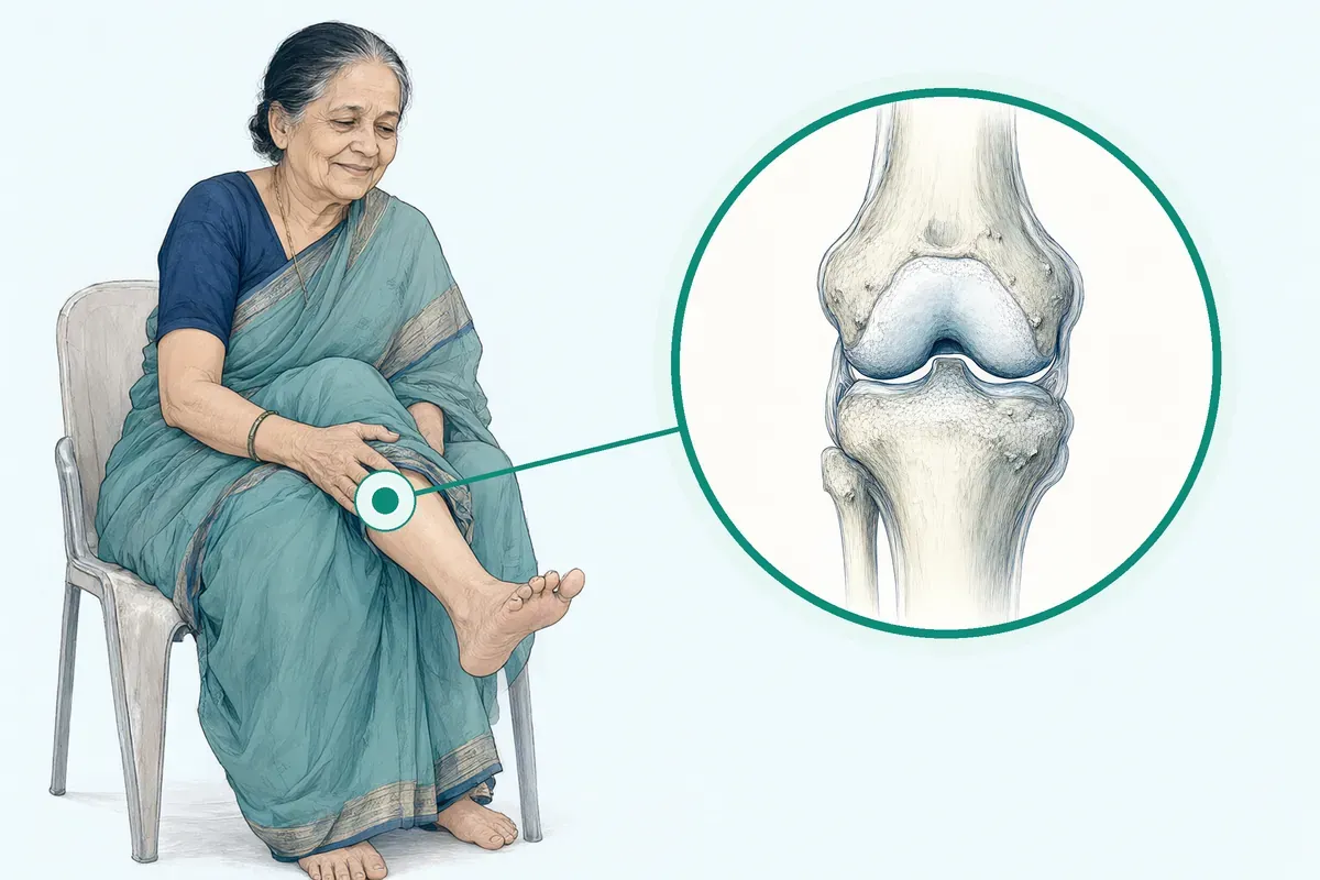
Osteoarthritis can involve almost any joint but typically affects the hands, knees, hips, and feet.
Why Jaipur Hospital
You may worry about worsening knee pain, prolonged time off work, and navigating surgical decisions.
Your care plan
Your first steps with us
- 1 Book an OPD consultation with our orthopaedic surgical team.
- 2 Review your joint scans and diagnostic tests in-house.
- 3 Discuss non-surgical therapies or surgical joint preservation plans.
- 4 Complete guided rehabilitation and scheduled follow-up visits.
How joint preservation care is planned
Your surgeon will discuss whether medical management, injections, or corrective bone procedures are suitable for your joint condition. Care typically ranges from daycare injections to a two-night stay for corrective bone surgery.
-
Intra-articular Injections
Your surgeon will discuss options such as hyaluronic acid, PRP, or corticosteroid injections into the joint.
-
Arthroscopic Debridement
Your surgeon will discuss keyhole joint cleaning to provide mechanical symptom relief for early osteoarthritis.
-
High Tibial Osteotomy (HTO)
Your surgeon will discuss realigning the shinbone to shift body weight away from the damaged medial compartment.
-
Cartilage Restoration (ACI/Microfracture)
Your surgeon will discuss targeted techniques to address focal chondral cartilage defects in younger individuals.
Why patients trust us
- 1991 Serving Jaipur for 35 years
- NABH Accredited hospital
- 5 Orthopaedics consultants
- 24×7 Emergency, ICU and ambulance
- 25 Cashless schemes and insurers
Clinical findings on joint therapies
-
2 weeks
Patients using capsaicin cream for joint pain typically experience some pain relief within the first two weeks.
-
13 weeks
A meta-analysis found corticosteroid injections reduced pain and improved function, with differences gradually diminishing after thirteen weeks.
-
33–50%
Studies show that in osteoarthritic knees, natural hyaluronic acid concentration decreases by 33 to 50 percent.
If it is left untreated
Osteoarthritis is a degenerative condition characterised by progressive cartilage degradation, subchondral damage, and bone remodelling. Leaving joint issues unaddressed can lead to chronic pain and functional limitations in daily activities. Persons with advanced symptoms and structural damage are candidates for total joint replacement.
Life after joint preservation treatment
Joint preservation aims to support joint mechanics and manage discomfort so you can manage routine daily tasks with greater ease.
-
Smoother everyday movement
Most people notice improved joint movement during regular daily walking and routine household activities.
-
Clear activity guidance
You typically receive structured exercise advice and physiotherapy plans to strengthen supporting muscles safely.
-
Planned joint monitoring
Regular follow-up visits help track joint alignment and cartilage stability over time.
Frequently asked questions
Questions patients ask about arthritis and joint preservation
Which problems are assessed under arthritis and joint preservation?
In short
Jaipur Hospital assesses early to moderate osteoarthritis of the knee, hip, and other joints under its arthritis and joint preservation service. The team evaluates cartilage wear, joint alignment, and mechanical symptoms such as catching or giving way. The goal is to preserve the natural joint and delay or avoid replacement surgery.
In detail
The service covers intra-articular injections with hyaluronic acid, PRP, or corticosteroids for inflammatory or mechanical pain. Arthroscopic debridement may relieve mechanical symptoms in early osteoarthritis. High tibial osteotomy realigns the leg to shift load off the damaged medial compartment.
Cartilage restoration techniques such as microfracture or autologous chondrocyte implantation address focal chondral defects in younger patients. Evidence shows biomechanical and inflammatory origins drive osteoarthritis, and obesity worsens the condition. Weight loss combined with exercise reduces knee compressive forces and inflammatory markers more than exercise alone.
A common belief
Many people assume joint pain means immediate replacement.
What actually happens
In fact, guidelines recommend exercise, weight management, injections, and realignment surgery first. These can delay replacement for years in the right patients.
Your next step at Jaipur Hospital
Your surgeon will review your history, examine the joint, and order weight-bearing X-rays or MRI if needed. A care plan is usually discussed at the first or second visit. Non-surgical options start promptly; osteotomy requires 6 to 12 weeks recovery.
How will the doctor decide which tests I need?
In short
The doctor at Jaipur Hospital selects tests based on your symptoms, examination findings and suspected arthritis stage. Weight-bearing X-rays are standard for all patients to assess joint space, osteophytes and alignment. MRI is added when bone marrow lesions, cartilage defects or soft tissue pathology need clarification. Blood tests may be ordered if inflammatory arthritis or infection is suspected.
In detail
Radiographic indicators such as marginal osteophytes and joint space narrowing are primary diagnostic tools. Several physical examination findings are useful diagnostically, including bony enlargement in knee osteoarthritis and pain elicited with internal hip rotation in hip osteoarthritis.
Bone marrow lesions, an alteration of bone marrow visible on T2-weighted MRI, are commonly identified in osteoarthritic knees and indicate higher progression risk.
If the surgeon suspects an inflammatory phenotype with stiffness, swelling and effusion, blood tests such as ESR, CRP or rheumatoid factor may be requested.
The choice of imaging and labs is tailored to the clinical question: X-rays for structure and alignment, MRI for cartilage and marrow, bloods for systemic inflammation.
A common belief
Patients often expect a full panel of tests.
What actually happens
In reality, tests are targeted: X-rays show structure and alignment, MRI shows cartilage and bone marrow lesions, bloods rule out inflammatory causes.
Your next step at Jaipur Hospital
Tests are usually done on the same day or within a few days using the in-house laboratory and radiology. Results are reviewed at a follow-up visit or by phone. The surgeon then finalises your treatment plan.
How soon should I expect improvement after the chosen arthritis treatment?
In short
At Jaipur Hospital, the timeline for improvement depends on the specific treatment you receive. Intra-articular injections such as corticosteroids often ease pain within a few days, while hyaluronic acid or PRP may take two to four weeks for full effect. For high tibial osteotomy, meaningful functional improvement typically appears over six to twelve weeks as the bone heals and rehabilitation progresses.
In detail
Evidence indicates that corticosteroid injections work quickly and can ease pain for several weeks or months, though the benefit gradually diminishes after up to thirteen weeks.
Hyaluronic acid injections are associated with symptom improvement beyond twelve weeks and have a more favourable safety profile than repeated corticosteroids. For osteotomy, the bone cut requires time to unite; protected weight-bearing and physiotherapy are essential during the first six weeks.
Studies on diet and exercise programmes show that combined interventions reduce pain and improve function more than either alone at eighteen months. Capsaicin cream may require up to a month for full effect. Your surgeon will set realistic expectations based on the technique chosen and your baseline joint health.
A common belief
Patients often expect instant relief from all treatments.
What actually happens
In reality, joint-preserving surgery needs a structured rehabilitation period, while injections vary in onset and duration.
Your next step at Jaipur Hospital
After injections, you can usually resume light activities the same day and return to normal routine within days. After osteotomy, you will use walking aids for several weeks and attend supervised physiotherapy. Follow-up visits are scheduled at two weeks, six weeks, and three months to monitor healing.
Is my joint pain caused by wear, inflammation, crystals, or another problem?
In short
At Jaipur Hospital, your surgeon determines the cause of joint pain through history, examination, and imaging. Osteoarthritis is a degenerative condition characterised by progressive cartilage degradation, subchondral damage, and bone remodelling with biomechanical and inflammatory origins. Inflammatory phenotypes show stiffness, swelling, and effusion. Crystal arthritis such as gout or pseudogout is identified by crystal analysis of joint fluid. Other causes include infection, avascular necrosis, or referred pain.
In detail
Evidence shows that knee osteoarthritis has biomechanical and inflammatory origins. Obesity exacerbates knee osteoarthritis by increasing joint load and systemic inflammation. Radiographic indicators include marginal osteophytes and joint space narrowing.
Bone marrow lesions on MRI are commonly identified in osteoarthritic knees and indicate higher progression risk. Patients with an inflammatory phenotype of osteoarthritis, characterised by stiffness, joint swelling, and effusion, are more likely to respond to corticosteroid injections compared to hyaluronic acid.
Physical examination findings such as bony enlargement in knee osteoarthritis and pain elicited with internal hip rotation in hip osteoarthritis guide diagnosis. Joint aspiration with crystal analysis distinguishes crystal arthropathy. Blood tests for inflammatory markers, uric acid, and infection help differentiate causes.
A common belief
Patients often assume all joint pain is simple wear and tear.
What actually happens
In reality, inflammatory phenotypes, crystal deposits, or other pathologies change the treatment approach entirely.
Your next step at Jaipur Hospital
Your surgeon will take a detailed history, examine the joint, and order appropriate imaging and laboratory tests. X-rays assess joint space and alignment. MRI evaluates cartilage, bone marrow, and soft tissues. Joint fluid analysis confirms or excludes crystals and infection. Blood tests screen for inflammatory or metabolic causes. Based on findings, a targeted treatment plan is developed.
How can I decide whether to seek advice about osteoarthritis management?
In short
Seek advice at Jaipur Hospital if joint pain, stiffness or swelling limits your walking, daily tasks or sleep. Early assessment helps preserve the joint. The hospital's orthopaedic team evaluates symptoms, examines the joint and may order X-rays or MRI to confirm osteoarthritis and grade its severity before suggesting a plan.
In detail
Osteoarthritis involves cartilage degradation, bone remodelling and synovial inflammation leading to pain, stiffness and functional limitation. Risk factors include age, female sex, obesity, genetics and previous joint injury. Being overweight doubles the odds of knee pain; obesity raises it 2.6 times.
Prior knee injury nearly triples the risk. NICE and OARSI guidelines recommend core non-surgical management for all: land or water exercise, strength training, weight management if overweight, education and self-management. Topical NSAIDs are effective for knee and hand osteoarthritis.
Oral NSAIDs need a proton pump inhibitor to protect the stomach. Intra-articular corticosteroids suit inflammatory phenotypes with effusion. Hyaluronic acid may help beyond 12 weeks. Duloxetine has demonstrated efficacy. Opioids are not superior to non-opioids for pain-related function and carry more adverse effects. Surgery is considered only when other treatments fail or daily life is seriously affected.
A common belief
People often wait until the joint is very stiff or deformed.
What actually happens
By then, preservation surgery may no longer be possible and replacement becomes the only choice. Starting earlier lets you use exercise, weight loss, injections or osteotomy to delay replacement.
Your next step at Jaipur Hospital
If pain persists despite simple measures, or if you notice reduced walking distance, difficulty with stairs or night pain, book an OPD consultation. The surgeon will review imaging, discuss injection options, physiotherapy, bracing or joint-preserving surgery such as osteotomy.
Recovery from injections is immediate to days; osteotomy needs 6 to 12 weeks. Typical cost range is ₹5,000 to ₹1,50,000.
How will arthritis treatment affect my walking, movement, or independence?
In short
Arthritis treatment at Jaipur Hospital aims to reduce pain, improve joint stability, and keep you walking independently. Non-surgical options like exercise, weight management, and injections can restore comfortable movement for daily tasks. Joint-preserving surgery such as osteotomy realigns the leg to offload the worn compartment, extending the life of your own joint.
In detail
Exercise strengthens muscles around the joint, which reduces load on cartilage and improves balance. In studies, diet plus exercise lowered knee compressive forces and gave better pain and function scores than exercise alone. Intra-articular corticosteroids give short-term relief to enable movement.
Hyaluronic acid and PRP may improve function for months in early arthritis. High tibial osteotomy shifts weight off the damaged medial compartment, allowing return to active walking over 6 to 12 weeks recovery. Cartilage restoration targets focal defects in younger patients.
The goal across all treatments is to maintain or regain the ability to walk, climb stairs, and perform self-care without assistance.
A common belief
Some fear treatment means long bed rest.
What actually happens
Most non-surgical options let you move straight away. Even after osteotomy, you start walking with support in days. The real risk to independence is doing nothing while the joint stiffens and weakens.
Your next step at Jaipur Hospital
After assessment, your surgeon outlines expected timelines. Injections allow immediate weight-bearing. Osteotomy needs crutches for 4 to 6 weeks, then progressive physiotherapy. Most patients resume household walking by 8 weeks and community distances by 12 weeks. A home exercise programme continues for months. Regular follow-up tracks joint health.
Which symptoms should prompt me to see a specialist?
In short
You should see a specialist at Jaipur Hospital if you have persistent joint pain, stiffness lasting more than 30 minutes in the morning, swelling, reduced range of motion, or a feeling of the joint giving way. Pain that limits walking, climbing stairs, or sleep also warrants review. Early assessment helps preserve the joint.
In detail
Osteoarthritis typically affects hands, knees, hips, and feet, causing pain, stiffness, functional limitation, and loss of valued activities. Risk factors include age, female sex, obesity, genetics, and previous joint injury. Being overweight doubles the odds of knee pain onset; obesity more than doubles it.
Previous knee injury nearly triples the risk. Guidelines recommend core management for all: exercise, weight loss if appropriate, and education, complemented by topical or oral NSAIDs where safe. Intra-articular corticosteroids give short-term relief. Hyaluronic acid may improve symptoms beyond 12 weeks. Duloxetine has shown efficacy. Opioids are not superior to non-opioid options and carry more adverse effects.
A common belief
People often wait until pain is severe.
What actually happens
Guidelines show early intervention with exercise, weight loss, and targeted injections preserves the joint longer. Delaying can mean more damage and fewer preservation options.
Your next step at Jaipur Hospital
At your visit, the surgeon will ask about symptom duration, impact on daily tasks, and previous treatments. Weight-bearing X-rays and sometimes MRI are ordered. A treatment plan is usually outlined within one to two visits. Non-surgical therapies can start immediately.
How will the results change my care plan?
In short
Imaging and blood results at Jaipur Hospital directly shape your preservation plan. X-ray joint space narrowing and alignment guide whether injection, debridement, or osteotomy is appropriate. MRI cartilage defects may indicate microfracture or autologous chondrocyte implantation. Inflammatory markers may shift care toward rheumatology input. Bone marrow lesions signal higher progression risk.
In detail
Evidence shows patients with bone marrow lesions are nine times more likely to progress to total knee replacement within three years. Hyaluronic acid concentration in normal knees is 2.5 to 4.0 mg/ml but drops 33 to 50 percent in osteoarthritis.
The OARSI guidelines classify treatments as appropriate for all, appropriate for specific sub-phenotypes, uncertain, or not appropriate. For example, intra-articular corticosteroids have a 1B high-consensus recommendation for short-term relief. Hyaluronic acid shows benefit beyond 12 weeks. High tibial osteotomy realigns load for medial compartment disease. Your surgeon matches the evidence to your imaging phenotype.
A common belief
People think all osteoarthritis gets the same treatment.
What actually happens
Guidelines define sub-phenotypes: inflammatory, mechanical, structural. Each has different appropriate therapies. Your imaging phenotype decides if you get corticosteroids, hyaluronic acid, realignment surgery, or cartilage restoration. One size does not fit all.
Your next step at Jaipur Hospital
After results are available, usually within a week, the surgeon explains the findings and revises the plan. Non-surgical treatments start promptly. Surgical options such as osteotomy need pre-operative assessment and scheduling. Recovery for osteotomy is 6 to 12 weeks.
What should I do if arthritis symptoms persist or worsen after treatment?
In short
If your arthritis symptoms persist or worsen after treatment at Jaipur Hospital, contact your surgical team promptly. Do not wait for the next scheduled visit if pain increases suddenly, swelling appears, or the joint feels unstable. Early review allows timely adjustment of medicines, physiotherapy, or further investigation such as repeat imaging.
In detail
Persistent pain after injections may indicate that the inflammation pattern or structural damage needs a different approach; for example, corticosteroid effects can wane by 13 weeks, and hyaluronic acid may not suit all phenotypes.
Worsening mechanical symptoms like locking or giving way after debridement could suggest progression of cartilage loss or new meniscal pathology. After osteotomy, increasing pain or inability to bear weight may signal delayed union or hardware issues.
Evidence notes that patients with bone marrow lesions are nine times more likely to progress to joint replacement within three years, so new or changing symptoms warrant re-evaluation.
The OARSI guidelines emphasise individualised management based on clinical sub-phenotypes, meaning a change in symptoms may shift the recommended treatment tier. Your surgeon will assess whether to escalate non-surgical care, repeat an injection, or consider the next surgical option.
A common belief
Many patients hope symptoms will settle on their own or keep taking painkillers.
What actually happens
But worsening mechanical symptoms can mean the joint is deteriorating faster, and delayed review may limit joint-preserving options. Guidelines stress re-assessment when the clinical picture changes.
Your next step at Jaipur Hospital
Keep a symptom diary noting pain level, swelling, and activity triggers. Call the hospital if you develop fever, redness, or sudden severe pain, which could indicate infection. For gradual worsening, request an earlier OPD appointment.
How do symptoms and scan findings influence the arthritis treatment plan?
In short
At Jaipur Hospital, your treatment plan is shaped by the combination of your symptoms, physical examination, and imaging findings. Pain severity, functional limitation, mechanical symptoms like locking or giving way, and the presence of swelling guide the urgency and type of intervention. X-rays show joint space narrowing, osteophytes, and alignment. MRI reveals cartilage defects, bone marrow lesions, and meniscal pathology. These findings determine whether non-surgical care, injections, joint-preserving surgery, or replacement is appropriate.
In detail
Evidence shows that radiographic indicators include marginal osteophytes and joint space narrowing. Bone marrow lesions on MRI are commonly identified in osteoarthritic knees; patients with bone marrow lesions are nine times more likely to progress to total knee arthroplasty within three years.
Physical examination findings such as bony enlargement in knee osteoarthritis and pain elicited with internal hip rotation in hip osteoarthritis are useful diagnostically. The inflammatory phenotype with stiffness, swelling, and effusion predicts better response to corticosteroid injections.
Focal chondral defects in young patients may suit cartilage restoration. Varus malalignment with medial compartment wear may indicate high tibial osteotomy. Advanced structural damage with persistent symptoms despite non-surgical care leads to joint replacement discussion. The multidisciplinary team integrates all data to match the intervention to your disease stage and phenotype.
A common belief
Patients often focus only on pain severity.
What actually happens
In fact, a meniscal tear on MRI without mechanical symptoms may not need surgery, while bone marrow lesions signal higher progression risk even with moderate pain.
Your next step at Jaipur Hospital
Bring all previous imaging and reports to your consultation. Your surgeon will correlate your symptom pattern with scan findings and explain which findings are driving the recommendation. Repeat imaging may be needed if prior scans are outdated. Treatment options, expected outcomes, and timelines will be discussed.
FAQ library
Questions and answers about arthritis and joint preservation
When you contact us
Consult our orthopaedic team in Jaipur
Contact Jaipur Hospital to schedule your orthopaedic OPD consultation and have your joint symptoms assessed.
- A consultation with an orthopaedic surgeon in the OPD
- Your scans and reports reviewed, with tests done in-house if you need them (laboratory, X-ray and imaging)
- Your options explained in plain words, including whether you need surgery at all
- A cost estimate before you decide
- Help with cashless treatment under RGHS, MAA Yojana, CGHS and empanelled insurers and TPAs
- Follow-up visits after your treatment
Overview
Arthritis and Joint Preservation: what to know
What is osteoarthritis and how joint preservation works
Osteoarthritis is a common joint condition that causes ongoing pain and difficulty with daily movement. It is characterised by progressive breakdown of cartilage, changes in the underlying bone, and alterations in the joint lining, ligaments, muscles, and surrounding fat. Over time, these structural changes can lead to joint dysfunction, morning stiffness, and difficulty walking or participating in daily activities. Knee osteoarthritis has both mechanical strain and inflammatory origins.
While osteoarthritis can involve almost any joint, it typically affects your knees, hips, hands, and feet. In a healthy knee, the concentration of natural hyaluronic acid ranges from 2.5 to 4.0 mg/ml, but in an osteoarthritic joint, this fluid decreases by 33 to 50 percent. The condition does not necessarily get worse for every person.
Joint preservation focuses on medical care, physical therapy, and joint-preserving procedures to protect your natural bone and cartilage before advanced damage occurs.
Symptoms of arthritis and warning signs that need urgent care
- Sudden severe swelling
- Rapid pain worsening
- Cannot bear weight
- Acute knee locking
Common symptoms of osteoarthritis include:
- Joint pain
- Stiffness
- Reduced movement
When your joints feel stiff or sore, you might worry that staying active will cause further harm, but careful movement is an essential part of care. Some individuals also notice visible bony enlargement around the affected joints.
In some people, osteoarthritis shows an inflammatory pattern marked by significant stiffness, noticeable swelling, and joint effusion. If you experience sudden, severe joint swelling, rapid worsening of pain, inability to bear weight, or acute mechanical locking of your knee, you should seek urgent evaluation the same day in our OPD or emergency department.
Causes of arthritis and who is at higher risk
- 24.6 percent New knee pain from overweight
- 5.1 percent New knee pain from past injury
- 33 percent Symptomatic knee OA over 75
Osteoarthritis develops from a combination of mechanical loading and joint inflammation. Being overweight places extra strain on weight-bearing joints such as your knees and hips, which often makes joint breakdown worse. In statistical analyses of new knee pain onset, 24.6 percent of cases were related to being overweight or having obesity, while 5.1 percent were linked to previous knee injuries.
Key risk factors include:
- Older age
- Female sex
- Genetics
- Obesity
- Major joint injuries
Among people older than 75 years, 33 percent have symptomatic and radiographic knee osteoarthritis. Research indicates that knee pain onset is significantly higher in females and in individuals with previous joint trauma. In contrast, studies show that smoking is not a statistically significant risk factor for the condition.
How arthritis is diagnosed: examination and tests at Jaipur Hospital
- Physical examination Check for bony enlargement and hip rotation pain
- Standing X-rays Look for bone spurs and joint space narrowing
- MRI scan Detect bone marrow lesions on T2-weighted images
Our clinical team assesses joint issues using a thorough physical examination and diagnostic imaging. During an OPD examination, physical indicators such as bony enlargement around the knee or pain provoked by internal hip rotation provide important diagnostic clues.
Your evaluation typically includes standing X-rays to check for bone spurs and joint space narrowing. Magnetic resonance imaging may also be used to detect bone marrow lesions, which are alterations in the marrow tissue visible on T2-weighted MRI scans.
Identifying these marrow changes is clinically significant, as study data show patients with bone marrow lesions are nine times more likely to progress to total knee replacement within three years compared to those without them.
Treatment options without surgery and when surgery is advised
Intra-articular Injections
- Hyaluronic acid, PRP, corticosteroids
Arthroscopic Debridement
- Clean debris, smooth surfaces
High Tibial Osteotomy
- Realign tibia, shift weight
Cartilage Restoration
- ACI or microfracture
Non-surgical care is the primary approach for early to moderate osteoarthritis.
Core management includes:
- Regular exercise
- Weight management
- Education
Land-based or water-based exercises strengthen muscles around your joints to help improve daily function and reduce discomfort. In an 18-month trial of overweight and obese adults with knee osteoarthritis, combining dietary weight loss with exercise lowered knee compressive forces and inflammatory markers like IL-6 while delivering better pain relief and physical function than exercise alone.
Medicines may be used alongside physical routines. Your doctor may suggest topical non-steroidal anti-inflammatory drugs, which can be bought over the counter at a chemist and applied directly to relieve pain and swelling in knees or hands.
Oral NSAIDs are an option, but because they can irritate your stomach lining, a chemist or doctor typically provides a proton pump inhibitor to protect your stomach. Capsaicin cream made from chillies can ease hand or knee pain by blocking local nerve signals, though it can cause a temporary burning sensation on the skin.
Assistive devices also reduce strain on affected joints. Shock-absorbing footwear, shoe insoles, knee braces, and splints help distribute weight evenly. If walking is difficult, using a walking stick on the side opposite your painful leg helps absorb body weight. Applying hot-water bottles or cold packs to your joint can also relieve symptoms.
When oral treatments and exercise do not provide sufficient relief, intra-articular injections are considered. These procedures typically take 30 to 60 minutes per session under local anaesthesia or no anaesthesia. Corticosteroid injections act quickly to reduce pain for several weeks or months, particularly in knees with swelling and inflammation.
Hyaluronic acid injections help restore joint lubrication, with symptom benefits observed beyond 12 weeks. Other injection techniques include platelet-rich plasma, ozone therapy, and mesenchymal stem cells.
Joint-preserving surgery is considered when non-surgical therapies have not worked and daily tasks remain severely limited. Common surgical options include:
- Intra-articular Injections: Delivering hyaluronic acid, PRP, or corticosteroids into the joint space to relieve pain and improve movement.
- Arthroscopic Debridement: Cleaning mechanical debris and smoothing joint surfaces for early osteoarthritis symptoms.
- High Tibial Osteotomy (HTO): Realigning the tibia bone to shift body weight away from the damaged medial compartment of the knee.
- Cartilage Restoration (ACI/Microfracture): Repairing isolated, focal cartilage defects in younger active patients.
Your surgeon will advise which option suits your joint condition. Recovery from injections ranges from immediate to a few days, while recovery following an osteotomy typically takes 6 to 12 weeks. Hospital stays usually range from daycare to 2 nights if an osteotomy is performed.
Typical costs for these procedures range from ₹5,000 to ₹1,50,000 depending on the specific procedure, hospital stay, and necessary diagnostic tests.
Preparing for arthritis and joint preservation procedures
Do
- Bring medical files and reports
- Bring government health scheme cards
- Have an attendant accompany you
Avoid
- Do not ignore fasting instructions
When you prepare for arthritis treatment or joint preservation surgery at Jaipur Hospital, your medical team will first review your overall health.
Diagnostic tests contribute to the total planning and cost, including:
- Blood tests
- Blood sugar tests
- Kidney function tests
- Liver function tests
These routine checks help our surgical team confirm that you are fit for your procedure.
You should talk to your doctor about all medicines you currently take. The type of pain medicine recommended for you depends on your pain levels and any other health conditions you have. If you take low-dose aspirin, ask your doctor whether you should use an NSAID.
Non-steroidal anti-inflammatory tablets may not be suitable if you have asthma, a stomach ulcer, angina, or a past history of heart attack or stroke. If you are given an oral NSAID, your doctor will usually also advise a proton pump inhibitor to protect your stomach lining against stomach acid. If you are prescribed opioids, a laxative may be added to manage constipation.
For joint procedures, your care team will give you clear fasting instructions before you arrive at the hospital. On the day of your appointment, remember to bring your previous medical files, test reports, prescription slips, and government health scheme cards. Having an attendant accompany you to the hospital helps with admission formalities and your return home.
How arthritis and joint preservation procedures are done
- Anaesthesia and sterilisation Local or general anaesthesia, then antibacterial skin prep
- Incision and guide wire Cut skin and place wire to mark bone section
- Bone cut and realignment Saw cuts bone, damaged section removed, gap closed
- Fixation Pins, rods, screws or plates hold bone while it heals
Jaipur Hospital provides non-surgical and surgical options for early to moderate arthritis. Sessions typically take 30 to 60 minutes. Treatments are usually carried out under local anaesthesia or with no anaesthesia. You may receive a local anaesthetic first to numb the joint area and reduce discomfort. Injections are given directly into the affected joint.
Your surgeon will advise which technique suits your joint condition. Intra-articular injections are one common option. These include hyaluronic acid, platelet-rich plasma (PRP), or corticosteroids. Corticosteroids contain synthetic cortisol and are used for painful joint problems. Platelet-rich plasma is prepared from your own blood and contains a higher concentration of platelets than ordinary blood. Both leukocyte-rich and leukocyte-poor PRP products are available.
Arthroscopic debridement is another option used to provide mechanical symptom relief in early osteoarthritis. For young patients with focal cartilage defects, cartilage restoration options include autologous chondrocyte implantation (ACI) and microfracture.
High tibial osteotomy is a joint-preserving surgery that realigns the leg for medial compartment knee osteoarthritis. During a knee osteotomy, the doctor cuts and reshapes the bones beneath the kneecap. Following anaesthesia, the surgical site is sterilised with an antibacterial solution. An incision is made, and doctors use a guide wire to mark the bone section for removal.
A surgical saw is used to cut into the bone. The damaged bone section is excised, and the gap is closed to align the edges. In some procedures, doctors insert a bone graft where bone was taken out. Pins, rods, screws, or plates are then fixed to hold the bone together while it heals. This shifts weight away from the damaged side of your knee.
Risks and possible complications of joint preservation treatments
- NSAIDs
- Can damage stomach lining
- Opioids
- Drowsiness, nausea, constipation; limited to short-term use
- Capsaicin cream
- Burning sensation on skin; avoid sensitive areas
- Ozone injections
- Minimal infection risk as ozone acts against microbes
All medical and surgical procedures carry potential risks. Surgery cannot be promised to clear your symptoms entirely, and you may still experience pain and stiffness. In studies, knee osteotomy relieves symptoms, though some patients may still need knee replacement surgery later.
When treatments target nerves, pain can return as the nerves grow back, meaning the procedure may need to be repeated.
Medicines used for arthritis also have documented side effects. Non-steroidal anti-inflammatory drugs can break down the protective stomach lining against digestive acid. Opioid painkillers can cause side effects such as drowsiness, nausea, and constipation, which is why doctors typically limit them to short-term use.
In a 12-month clinical trial, adverse medication-related symptoms were significantly more common in patients taking opioids (mean 1.8 symptoms) than in those taking non-opioid medicines (mean 0.9 symptoms). Capsaicin cream, made from chillies, can cause a burning sensation on the skin, and touching sensitive areas can cause sharp pain for several hours. With ozone gas injections, the risk of infection is minimal because ozone acts against bacteria, fungi, and viruses.
Physical therapy also requires care. In studies and clinical guidance, doing too much exercise too quickly or choosing the wrong exercises carries a risk of damaging your joints.
Recovery and aftercare following joint preservation
Your recovery timeline depends on the specific procedure you have. After joint injections, recovery is typically immediate to a few days. For high tibial osteotomy, recovery typically takes 6 to 12 weeks. Your stay at Jaipur Hospital is typically daycare for minor treatments, or up to 2 nights if you undergo an osteotomy.
In the first days after treatment, rest and careful joint care are essential. Using hot or cold packs on your joints can help reduce pain and swelling. A hot-water bottle filled with warm or chilled water can be applied directly to the joint.
Reusable packs that are cooled in a freezer or warmed in a microwave work in the same way. If you use capsaicin cream, apply a pea-sised amount up to 4 times a day, keeping at least 4 hours between applications. Never apply it to broken or inflamed skin.
Always wash your hands after applying it, and avoid hot baths before or after application. Some pain relief from the cream is usually noticed within 2 weeks, while full effect can take up to a month.
During later weeks, physical therapy helps you rebuild movement and strength. A physiotherapist can use manual therapy to stretch and massage tissues to keep your joint mobile. Assistive devices can make walking and daily work easier. Using a walking stick on the side opposite your painful leg helps take weight off the joint. Shock-absorbing footwear, shoe insoles, splints, and leg braces also help spread weight evenly across your limbs.
Contact Jaipur Hospital promptly if you develop a high fever, sudden severe calf or joint pain, increasing redness, or fluid leakage from your surgical wound.
Results and long-term outcomes in clinical studies
Clinical trials show that joint preservation and medical care can reduce joint pain and support daily physical function. In a large Cochrane meta-analysis of 27 randomised controlled trials, corticosteroid injections reduced pain and improved joint function compared to control treatments, though the difference gradually faded by 13 weeks of follow-up.
Another study noted that intra-articular hyaluronic acid injections provide symptom improvement beyond 12 weeks with a more favourable safety record than repeated steroid shots. In clinical trials of radiofrequency denervation for knee osteoarthritis, pain reduction lasted for up to 2 years.
Long-term trials also show the value of guided lifestyle changes. In an 18-month clinical trial of 454 overweight and obese adults with knee osteoarthritis, participants in a combined diet and exercise group achieved an average weight loss of 10.6 kg (11.4%), compared to 8.9 kg (9.5%) in the diet-only group and 1.8 kg (2.0%) in the exercise-only group.
The combined diet and exercise group experienced less joint pain (score 3.6) and better physical function (score 14.1) than those who did diet alone (pain 4.8; function 18.4) or exercise alone (pain 4.7). Inflammatory IL-6 markers and compressive knee forces were also lower in groups that altered their diet.
Regarding pain medicines, a 12-month clinical trial found that opioid therapy was not superior to non-opioid treatments for pain-related function, while pain intensity was significantly better in the non-opioid group.
Cost of joint preservation and insurance in Jaipur
The typical price range for arthritis and joint preservation procedures in India is ₹5,000 to ₹1,50,000. For an individual estimate, please consult our team at Jaipur Hospital. Published hospital data in India shows that an osteotomy typically averages from Rs. 80,000 to Rs. 2,50,000. Across different cities, osteotomy costs range from Rs. 80,000 to Rs.
1,80,000 in Raipur, Indore, and Aurangabad; Rs. 1,00,000 to Rs. 1,80,000 in Visakhapatnam and Nagpur; Rs. 1,00,000 to Rs. 2,20,000 in Bhubaneshwar; and Rs. 1,20,000 to Rs. 2,50,000 in Hyderabad.
Several factors determine your total bill. These include doctor consultation fees, diagnostic blood and organ function tests, the specific type of surgery performed, and the severity of your joint damage.
Costs also depend on the surgical tools, implants, and machines needed during the operation, the type of hospital chosen, the number of days you stay in a hospital room, and your post-operative medicines and follow-up visits.
Jaipur Hospital is a NABH-accredited multispeciality hospital established in February 1991. We accept government healthcare schemes including RGHS, MAA Yojana, and CGHS, alongside a panel of private insurers and TPAs. Please bring your scheme documents and insurance cards to our desk during admission so our team can help you verify your coverage.