

Orthopaedics · Jaipur Hospital
Fracture and Trauma Surgery in Jaipur
Broken bone from a fall or accident? We assess the injury and give you a clear treatment plan.
- Serving Jaipur since 1991 (35 years)
- NABH accredited
- 24×7 emergency & ICU
- Cashless: RGHS, MAA Yojana, CGHS + 22 insurers
- Operation time
- 1 to 4 hours (varies by injury)
- Anaesthesia
- General or regional anaesthesia
- Recovery
- Weight-bearing per fixation stabilityfull union in 3 to 6 months
- Price
- ₹50,000 to ₹3,00,000typical range in India; ask us for an estimate
Broken bone, disrupted life
A fracture stops you from moving, working, and caring for your family
Pain, swelling, and inability to bear weight make daily tasks impossible. Without proper fixation, the bone may heal in the wrong position, leading to long-term stiffness or limp.
-
Sudden pain and swelling
A fall or crash leaves you unable to put weight on the limb.
-
Fear of wrong healing
If bones are not aligned, they may knit together crookedly.
-
Loss of income
Time off work adds financial stress to physical recovery.
-
Complex injuries
Polytrauma needs coordinated care across specialties.
Fracture fixation aims to provide stability and promote healing.
Why Jaipur Hospital
You worry about the pain, the weeks off work, and whether the bone will knit back correctly.
Your care plan
Your first steps with us
- 1 Call or visit our 24x7 emergency department
- 2 Get X-rays and scans reviewed by orthopaedic surgeon
- 3 Undergo fixation surgery with appropriate anaesthesia
- 4 Follow your personalised rehab plan at home
How the operation is done
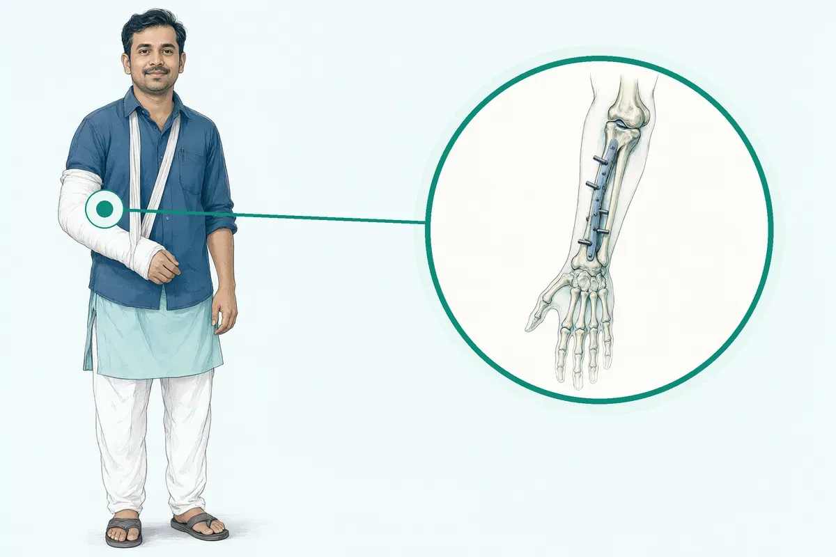
The surgeon realigns the bone fragments and secures them with implants. Anaesthesia is general or regional. Operation time is usually 1 to 4 hours. Hospital stay is typically 2 to 7 nights.
-
Open Reduction Internal Fixation (ORIF)
Plates, screws, or nails hold the fragments in correct position for stable healing.
-
Intramedullary Nailing
A rod inside the bone canal shares load, often used for thigh or shin fractures.
-
External Fixation
Pins and an outside frame stabilise severe open fractures until soft tissues settle.
-
Minimally Invasive Plate Osteosynthesis (MIPO)
Percutaneous plating through small cuts preserves soft tissue and blood supply.
Why patients trust us
- 1991 Serving Jaipur for 35 years
- NABH Accredited hospital
- 5 Orthopaedics consultants
- 24×7 Emergency, ICU and ambulance
- 25 Cashless schemes and insurers
What research shows about fracture fixation
-
about 9 in 10
Around 75% of people who had ORIF for a fractured femur had few or no long-term complications in one study.
-
3 to 6 months
Full bone union typically occurs within this period, though severe fractures may take a year or longer.
-
about 20 in 100
Reoperation within 24 months occurred in roughly 20% of hip fracture patients regardless of fixation type in a large trial.
If it is left untreated
Without surgery, severe fractures often heal in a deformed position, causing chronic pain, limp, arthritis, and higher risk of re-fracture.
Life after treatment
Most people regain movement and return to daily activities as the bone unites over months.
-
Walk without support
Weight-bearing progresses per surgeon advice; many transition off crutches within weeks.
-
Return to work
Average recovery for desk work is 3 to 4 weeks; heavy labour takes longer.
-
Metal stays in place
Plates and screws usually remain permanently and rarely cause issues.
-
Gradual strength return
Physiotherapy helps restore joint range and muscle power over 3 to 6 months.
Frequently asked questions
Questions patients ask about fracture and trauma surgery
Which problems are assessed under fracture and trauma surgery?
In short
Fracture and trauma surgery at Jaipur Hospital assesses broken bones, joint dislocations, and multiple injuries from accidents or falls. The surgical team evaluates the bone pattern, soft tissue damage, and whether the fracture is open or closed. Polytrauma patients are checked for chest, head, and abdominal injuries alongside the fractures.
In detail
The assessment follows a structured approach. In the emergency department, the team stabilises life-threatening injuries first, then uses X-rays, CT scans, or MRI to define the fracture pattern and associated injuries.
Evidence shows rib fractures are the most common thoracic injury in blunt trauma, while pneumothorax and haemothorax occur in a majority of polytrauma patients with chest trauma. The fracture pattern guides the choice between plating, nailing, or external fixation.
For femoral neck fractures, the pattern determines whether a sliding hip screw, cancellous screws, or a cephalomedullary nail offers the most stable construct.
Polytrauma patients often need ICU care; studies report a median ICU stay of three days and a median hospital stay of eleven days for those with chest injuries.
A common belief
People often think only the broken bone is treated.
What actually happens
In polytrauma, chest injuries like pneumothorax or haemothorax are common and must be managed first. The fracture pattern decides whether plates, nails, or external fixation is used.
Your next step at Jaipur Hospital
You will be seen in the 24×7 emergency department or the orthopaedic OPD. Bring any previous imaging and a list of medicines. The surgeon will explain the injury, the likely fixation method, and the expected hospital stay of two to seven nights.
How will the doctor decide which tests I need?
In short
The doctor at Jaipur Hospital decides tests based on the injury mechanism, the body part involved, and the clinical examination findings. X-rays are the first test for most fractures. CT scans are added for complex joints, pelvic injuries, or when the fracture pattern is unclear on X-ray. MRI may be used if ligament or soft tissue injury is suspected.
In detail
Test selection follows a stepwise logic. After stabilising the patient, the emergency team orders X-rays to confirm any fracture.
If the fracture involves a joint surface, the pelvis, or the spine, or if the X-ray does not fully explain the pain, a CT scan gives three-dimensional detail for surgical planning.
Evidence shows finite element analysis of CT data helps surgeons understand implant stress and bone strain around screws. MRI is reserved for suspected ligament tears, occult fractures, or spinal cord injury.
For polytrauma, additional tests include chest X-ray, ultrasound, or CT of the chest, abdomen, and head to rule out internal injuries.
Studies report that ninety percent of polytrauma patients with blunt chest trauma were treated conservatively, but imaging guided the decision for chest tube insertion in fifty-five percent. The surgeon orders only the tests that change the treatment plan.
A common belief
Patients sometimes ask for every scan.
What actually happens
Unnecessary tests add cost and delay without changing management. The surgeon orders imaging that defines the fracture pattern, joint involvement, or associated injuries that alter the surgical approach.
Your next step at Jaipur Hospital
You will have X-rays in the in-house radiology department, often on the same visit. CT or MRI may be scheduled the same day or the next. The surgeon reviews the images with you and finalises the plan.
How soon should I expect improvement after fracture fixation surgery?
In short
After fracture fixation surgery at Jaipur Hospital, the first one to three days are usually the most uncomfortable. Pain decreases steadily as the wound heals. Bone union typically takes three to six months, but functional improvement starts earlier depending on the fracture site and fixation stability.
In detail
Recovery follows a phased timeline. In the first few days, pain is managed with medication and elevation. Sutures are usually removed at two to three weeks. You can often bathe and gradually increase activity after suture removal.
Weight-bearing depends on fixation stability; your surgeon will guide you. Swelling in the foot and ankle may persist for three to six months. Full recovery is often reached by twelve months.
In studies, around 75 percent of people who had ORIF for a fractured femur had few or no long-term complications. The median hospital stay for polytrauma patients with chest injuries was eleven days, with ICU stay of three days.
Time off work varies by occupation, averaging three to four weeks for foot and ankle procedures if there are no complications.
- In studies, around 75 percent of people who had ORIF for a fractured femur had few or no long-term complications.
A common belief
People often expect to be back to normal in a few weeks.
What actually happens
In reality, swelling can last three to six months, and full recovery may take a year. Rushing weight-bearing before the fixation is ready can cause failure.
Your next step at Jaipur Hospital
Follow your surgeon's customised recovery plan. Attend physiotherapy sessions as advised. Keep the limb elevated and take prescribed pain medication. Do not smoke, as nicotine slows healing. Return for suture removal at two to three weeks.
How will the fracture be assessed for displacement, instability, or damage to nerves and blood vessels?
In short
At Jaipur Hospital, the emergency team stabilises you first, then uses imaging to assess the fracture. X-rays confirm the fracture pattern and displacement. CT scans provide three-dimensional detail for complex fractures. MRI evaluates soft tissue, nerve, and blood vessel injury. Clinical examination checks pulses, sensation, and movement.
In detail
Assessment follows a structured sequence. In the emergency room, the team treats life-threatening injuries first. After stabilisation, X-rays are taken to confirm fractures. The provider may also use MRI or CT to look for other injuries.
For polytrauma with blunt chest trauma, rib fractures are common, and pneumothorax or hemothorax are assessed. Clinical examination evaluates nerve function and vascular status. The fracture pattern determines the ideal implant type.
Finite element analysis is used in research to understand fixation strength, but clinical decisions rely on imaging and examination. Each patient is considered individually, taking into account all patient and implant factors.
A common belief
Some people think an X-ray is enough.
What actually happens
Complex fractures need CT for surgical planning. Nerve and vessel injuries can be missed without a careful clinical exam and sometimes MRI. Polytrauma patients need chest and abdomen checks too.
Your next step at Jaipur Hospital
Go to the 24×7 emergency department immediately after injury. The team will perform clinical examination and order X-rays, CT, or MRI as needed. Your surgeon will explain the findings and whether surgery is required.
How can I decide whether to seek advice about closed reduction and casting?
In short
You should seek advice about closed reduction and casting when a fracture is stable, well aligned, and likely to heal without surgery. Jaipur Hospital evaluates each injury with imaging and clinical examination to determine whether non-operative treatment is appropriate for your specific fracture pattern.
In detail
Closed reduction means the bone fragments are realigned without opening the skin, then held in place with a cast or splint.
This approach is typically chosen for fractures that are not displaced, involve the shaft of a long bone in a stable pattern, or occur in bones with good blood supply.
Evidence shows that stable fractures treated non-operatively can unite well, avoiding surgical risks such as infection, implant irritation, or anaesthesia complications.
However, if the fracture is displaced, angulated, involves a joint surface, or shows signs of instability on follow-up X-rays, surgical fixation may become necessary to restore alignment and function. The decision depends on fracture type, bone quality, your age, activity level, and any associated injuries. Your surgeon will review serial imaging to confirm the position is maintained during healing.
A common belief
Many people assume all fractures need plates or nails.
What actually happens
In reality, stable fractures often heal reliably without surgery, avoiding infection and implant problems. But if the position shifts in the cast, surgery may still be needed later.
Your next step at Jaipur Hospital
If you have a recent injury or a cast that feels loose, painful, or tight, attend the emergency department or orthopaedic OPD at Jaipur Hospital for assessment. Bring any previous X-rays.
The surgeon will examine you, order imaging if needed, and discuss whether closed treatment remains suitable or if fixation is advised.
How will treatment affect my walking, movement, or independence?
In short
Treatment aims to restore your walking, movement, and independence by achieving stable fracture union and early mobilisation. Jaipur Hospital plans fixation and rehabilitation to help you return to your usual activities as safely and quickly as your injury allows.
In detail
The impact on walking and independence depends on the fracture site, fixation stability, and your pre-injury function. Stable internal fixation with plates or nails often allows partial or full weight-bearing within days to weeks, guided by your surgeon.
Hip fracture fixation typically permits immediate or early weight-bearing with a walking aid. Pelvic and acetabular fractures may require protected weight-bearing for six to twelve weeks. Physiotherapy starts in hospital and continues after discharge to regain strength, range of motion, and gait confidence.
Evidence shows that early mobilisation reduces complications such as deep vein thrombosis, chest infection, and muscle wasting. Your surgeon and physiotherapist will give you a personalised weight-bearing and exercise plan. Assistive devices such as crutches, a walker, or a wheelchair may be needed temporarily.
A common belief
Many patients fear they will be bedridden for months.
What actually happens
In fact, modern fixation allows early mobilisation. Delaying movement causes stiffness, weakness, and blood clots. Following the prescribed weight-bearing plan is key to regaining independence.
Your next step at Jaipur Hospital
Before discharge, the physiotherapy team at Jaipur Hospital will teach you safe transfers, walking with aids, and home exercises. Follow-up visits assess healing and progress your weight-bearing. Most patients regain independence within three to six months, though complex injuries may take longer.
Which symptoms should prompt me to see a specialist?
In short
See a specialist at Jaipur Hospital if you have severe pain, swelling, deformity, inability to move or bear weight on a limb, or an open wound near a suspected fracture. Numbness, tingling, or colour change in the fingers or toes also needs urgent evaluation.
In detail
These symptoms suggest a broken bone, dislocation, or injury to nerves and blood vessels. Pain that does not settle with rest and simple analgesia, worsening swelling, or a limb that looks shortened or angulated are typical signs.
In polytrauma, breathlessness, chest pain, or confusion may indicate associated chest or head injuries. Early specialist review allows timely imaging and fixation, which reduces the risk of malunion, nonunion, or compartment syndrome. The evidence shows that delayed fixation in unstable fractures increases complication rates.
A common belief
Many people try to walk on a painful limb or wait for swelling to go down.
What actually happens
Delaying review can lead to poor alignment, nerve damage, or infection, especially in open fractures.
Your next step at Jaipur Hospital
Do not wait if you notice any of these signs. Attend the 24×7 emergency department or book an OPD appointment. The surgeon will examine you, order X-rays or CT, and advise on the need for surgery.
How will the results change my care plan?
In short
Imaging results at Jaipur Hospital directly shape the care plan by defining the fracture pattern, displacement, joint involvement, and any associated injuries. The surgeon uses this information to decide between non-operative treatment, open reduction internal fixation with plates or screws, intramedullary nailing, external fixation, or minimally invasive plating. The choice of implant and the surgical approach depend on the fracture geometry seen on the scans.
In detail
The fracture pattern is the major determinant of the ideal implant.
For example, evidence shows that in Pauwels type III femoral neck fractures a cephalomedullary nail is significantly stronger in axial loading, while in unstable basicervical patterns cannulated screws in a triangular configuration demonstrated a lower ultimate load to failure.
In subcapital or transervical patterns both cannulated screws and a sliding hip screw demonstrated no compromise in fixation strength. For long bone shafts, intramedullary nailing provides load-sharing fixation. Severe open fractures with soft tissue loss may need temporary external fixation before definitive plating.
CT scans also reveal comminution, articular step-off, and bone quality, which influence whether a minimally invasive approach is possible. Polytrauma imaging identifies chest, abdominal, or head injuries that may delay fracture surgery until the patient is stable.
The surgeon integrates all findings to create a plan that addresses the most urgent problems first and selects the fixation method that gives stable alignment for healing.
A common belief
Patients often think all fractures get the same plate.
What actually happens
In reality, a femoral neck fracture may need a nail, screws, or a sliding hip screw depending on the angle. A comminuted tibia shaft may need a nail, while an open fracture with skin loss needs an external fixator first. The imaging detail drives every decision.
Your next step at Jaipur Hospital
After the scans, the surgeon will explain the findings, the recommended fixation, the expected operation time of one to four hours, and the recovery milestones. You will receive a cost estimate in the typical range of fifty thousand to three lakh rupees.
What should I do if symptoms persist or worsen after fracture fixation surgery?
In short
If you notice increasing pain, swelling, redness, fever, or numbness after fracture fixation surgery, contact your surgeon at Jaipur Hospital immediately. These signs may indicate infection, implant migration, nerve injury, or delayed healing. Early reporting allows prompt assessment and management.
In detail
Complications can arise after fixation. Infection after fracture fixation may result in functional loss. Deep infections involving the fixation are a reason for removal. Metal allergy can also require removal. Non-union means the bone fails to knit together, so fixation may be replaced.
Prominent fixation can protrude through the skin, increasing infection risk. Fixation that has migrated can be difficult to locate. Scars can become thickened and sensitive. Numbness from tiny nerve damage may improve or remain. Other risks include blood clots, chronic pain, and bleeding.
In rare cases, a re-fracture occurs during surgery. If you feel too much pain or worry about medication side effects, talk to your surgeon.
A common belief
Some patients wait weeks hoping symptoms settle.
What actually happens
Delaying can turn a manageable infection into a deep infection requiring implant removal, or miss a migrating screw that damages tissue. Early action prevents bigger problems.
Your next step at Jaipur Hospital
Call your surgeon or the hospital if you experience worsening pain, wound discharge, fever, new numbness, or inability to move the limb. Do not wait for the next scheduled visit. The 24×7 emergency department is available for urgent concerns.
Can the fracture heal with a cast or brace, or is surgery needed?
In short
Whether a fracture heals with a cast or needs surgery depends on the fracture type, displacement, and stability. Jaipur Hospital assesses each injury individually. Stable, non-displaced fractures often heal with immobilisation. Displaced, unstable, or open fractures usually require surgical fixation to restore alignment and prevent complications.
In detail
Evidence shows that severe fractures will not heal correctly without surgery. A bone that does not heal properly is more likely to break again. ORIF ensures the bone pieces are aligned properly. ORIF is usually very effective.
Success rates depend on which bone is fractured and the fracture pattern. Studies consistently find that ORIF is the preferred way to treat severe bone fractures that will not heal with only immobilisation.
Techniques include ORIF with plates and screws, intramedullary nailing for long bones, external fixation for severe open fractures, and MIPO for soft tissue preservation. The fracture pattern is the major determinant of the ideal implant. The surgeon considers patient factors, bone quality, and soft tissue condition before deciding.
A common belief
Patients sometimes prefer a cast to avoid surgery.
What actually happens
But evidence shows severe fractures will not heal correctly without fixation, and a poorly healed bone breaks again more easily. The fracture pattern, not patient preference, decides the treatment.
Your next step at Jaipur Hospital
After injury, attend the emergency department for X-rays. The surgeon will review the images and examine you. If the fracture is stable and aligned, a cast or brace may be advised with follow-up X-rays. If displaced or unstable, surgery will be discussed.
FAQ library
Questions and answers about fracture and trauma surgery
When you contact us
Get your fracture assessed today
Contact Jaipur Hospital for an orthopaedic OPD consultation, in-house scans, a clear explanation of options, a cost estimate, and help with cashless schemes.
- A consultation with an orthopaedic surgeon in the OPD
- Your scans and reports reviewed, with tests done in-house if you need them (laboratory, X-ray and imaging)
- Your options explained in plain words, including whether you need surgery at all
- A cost estimate before you decide
- Help with cashless treatment under RGHS, MAA Yojana, CGHS and empanelled insurers and TPAs
- Follow-up visits after your treatment
Overview
Fracture and Trauma Surgery: what to know
What fracture and trauma surgery is
- Simple fracture
- A crack in the bone
- Open fracture
- Bone pierces the skin
- Polytrauma
- Two or more serious injuries
A fracture is a broken bone. It happens when a force hits the bone that is stronger than the bone itself.
Common causes are:
- Falls
- Road traffic accidents
- Sports injuries
The break may be a simple crack or the bone may shatter into several pieces. In an open fracture the broken bone pierces the skin; this carries a higher risk of infection. Fracture fixation surgery aims to put the bone fragments back into their normal position and hold them there with metal implants so the bone can heal. The metalwork — plates, screws or rods — stays inside the body while the bone knits together.
Polytrauma means two or more serious injuries in different body regions at the same time. For example, a person may have a broken leg, a chest injury and a head injury after a high-speed crash. These patients need coordinated care from several specialties, often starting in the emergency department and continuing in the intensive care unit.
Symptoms of a fracture and when to seek urgent care
- Limb deformed, cold, pale or numb
- Open wound with bone visible
- Cannot move fingers or toes
- Pain not controlled by simple painkillers
- Difficulty breathing or chest pain
Typical signs of a fracture are:
- Pain that gets worse when you move or put weight on the injured part
- Swelling
- Bruising
- A visible deformity or shortening of the limb
You may hear a snap or grinding sound at the moment of injury. In an open fracture you will see bone or a deep wound with bleeding.
Go to the emergency department the same day if:
- The limb looks deformed, is cold, pale or numb
- There is an open wound with bone visible
- You cannot move the fingers or toes beyond the injury
- Pain is not controlled by simple painkillers
- You have difficulty breathing or chest pain after a fall or crash (this may mean a rib fracture or lung injury)
At Jaipur Hospital the 24×7 emergency department and ambulance service are ready to stabilise you and start treatment without delay.
Causes and people at higher risk
- Smoking or vaping
- Nicotine slows bone healing
- Poor nutrition
- Low vitamin C and D levels
- Uncontrolled diabetes
- Chronic illness affects healing
- Obesity
- Adds mechanical stress on bone
Most fractures result from trauma: falls from height, road traffic accidents, direct blows and sports injuries. Traffic accidents are the most common mechanism reported in large trauma series. In older people, even a minor fall can break a bone because the bone is thinner and weaker (osteoporosis).
Certain fracture patterns — such as unstable or osteoporotic fractures — carry a higher risk that the fixation will fail or the bone will not unite.
Risk factors that can slow healing or increase complications include:
- Smoking or vaping nicotine products
- Poor nutrition, especially low vitamin C and D levels
- Uncontrolled diabetes or other chronic illnesses
- Obesity, which adds mechanical stress on the healing bone
- Open fractures where the skin is broken, because bacteria can enter the wound
Your surgeon will ask about these factors before planning treatment.
How a fracture is diagnosed: examination and tests
- X-ray
- Shows bone outline and alignment
- MRI
- Checks soft tissue and ligament injury
When you arrive after an injury, the trauma team first checks your airway, breathing and circulation. Life-threatening injuries are treated before the fracture itself. Once you are stable, the doctor examines the injured area for swelling, deformity, open wounds and nerve or blood vessel function.
Imaging confirms the fracture and guides the surgical plan:
- X-rays are the first test; they show the bone outline, fracture pattern and alignment.
- CT scan gives detailed three-dimensional views, especially useful for complex joints, pelvis or spine fractures.
- MRI may be used when soft tissue, ligament or cartilage injury is suspected.
In polytrauma, chest X-ray or CT often shows associated injuries such as pneumothorax (air around the lung), hemothorax (blood around the lung) or pulmonary contusion (lung bruising). These findings help the team decide which surgery to do first and whether you need intensive care monitoring.
Non-surgical treatment and when surgery is advised
Do
- Bone fragments displaced, angulated or shortened
- Open fracture with bone through skin
- Joint surface stepped or gapped
- Multiple injuries needing early mobilisation
Not every fracture needs an operation. Stable fractures where the bone ends are well aligned can heal in a plaster cast, splint or brace. The doctor will check alignment with repeat X-rays over several weeks. In polytrauma, many chest injuries are managed without surgery — for example, 90% of blunt chest trauma patients in one large series were treated conservatively.
Surgery is advised when:
- The bone fragments are displaced, angulated or shortened so that the limb would heal in a poor position
- The fracture is open (bone through the skin) — urgent cleaning and fixation reduce infection risk
- The joint surface is stepped or gapped, which leads to early arthritis if not corrected
- The fracture is unstable and would not hold in a cast
- You have multiple injuries and early fixation allows you to mobilise sooner, reducing complications like pneumonia or blood clots
Common surgical methods include:
- Open reduction and internal fixation (ORIF) with plates and screws
- Intramedullary nailing for long bone shafts
- External fixation for severe open fractures as a temporary measure
- Minimally invasive plate osteosynthesis (MIPO) that preserves soft tissue
Your surgeon will explain which technique suits your fracture pattern and general condition.
Preparing for fracture and trauma surgery
Do
- Regular medicines
- List of allergies
- Comfortable loose clothing
- Attendant to stay after discharge
Before your operation, the surgical team will review your injuries and general health. You will usually need X-rays, and sometimes a CT scan or MRI, to confirm the fracture pattern and plan the fixation. Blood tests and an anaesthesia check are routine.
Tell your surgeon about every medicine you take, especially blood thinners, steroids, or supplements, so they can advise which to pause before surgery. You will be asked to fast for at least six hours before a general anaesthetic. If you are having regional anaesthesia, the anaesthesiologist may still give you medicine to make you sleepy.
Bring your regular medicines, a list of allergies, comfortable loose clothing, and an attendant who can stay with you after discharge. For polytrauma, the emergency team stabilises life‑threatening injuries first, then proceeds to fracture fixation once you are stable.
How fracture and trauma surgery is done
Open Reduction Internal Fixation
- ORIF
Rod inside long bone
- Intramedullary nailing
Pins and outside frame
- External fixation
Minimally invasive plates
- MIPO
On the day of surgery, you will be taken to the operating theatre and given either general anaesthesia (you sleep through the procedure) or regional anaesthesia (the area around the broken bone is numbed). The operation typically lasts one to four hours, depending on the bone and the severity of the break.
Your surgeon will choose the fixation method that suits your injury. Open Reduction Internal Fixation (ORIF) uses plates, screws, or nails to hold the fragments in place. Intramedullary nailing places a rod inside the hollow centre of a long bone for load‑sharing stability. External fixation uses pins and an outside frame, often as a temporary measure for severe open fractures.
Minimally Invasive Plate Osteosynthesis (MIPO) inserts plates through small incisions to preserve soft tissue. The surgeon makes an incision, realigns the bone fragments, applies the chosen implants, and closes the wound with stitches and a dressing. A plaster cast is not usually needed after internal fixation. Your surgeon will discuss which technique is appropriate for you before you sign the consent form.
Risks and possible complications of fracture and trauma surgery
All operations carry risks. Infection at the fixation site occurs in a small proportion of patients and can lead to further surgery or, in severe cases, loss of limb function. In one series of polytrauma patients, pneumonia was seen in about 13 in 100 people, sepsis in about 5 in 100, and multi‑organ failure in about 3 in 100.
Avascular necrosis (loss of blood supply to the bone end) was reported in 5 to 9 in 100 patients with certain hip fractures. Non‑union, where the bone fails to knit together, may require another operation. Implants can sometimes become prominent under the skin, causing discomfort or risk of infection.
Nerve injury may cause numbness that can improve with time or remain long‑term. Scars can thicken and stay sensitive. Blood clots, bleeding, and chronic pain are also recognised surgical risks. In rare cases, the bone can re‑fracture during fixation, requiring new implants. Smoking and nicotine use slow healing and raise complication rates. Your team will explain the risks that apply to your specific fracture.
Recovery and aftercare after fracture and trauma surgery
After the operation you spend a few hours in the recovery room while staff monitor your vital signs and pain. Most patients stay in hospital for two to seven nights; polytrauma patients may need ICU care. The first one to three days are usually the most uncomfortable.
You will be given a plan for pain relief that may include prescription medicine, NSAIDs if safe for you, and paracetamol. Keep the operated limb elevated as much as possible and move only for essential needs in the first few days.
Weight‑bearing depends on how stable the fixation is; your surgeon will tell you when and how much weight you can take. Stitches or clips are usually removed at two to three weeks. You can bathe once the wound is healed and gradually increase activity as comfort allows. A post‑operative shoe and crutches are often provided for lower‑limb injuries.
Physiotherapy starts early to maintain joint movement and muscle strength. Swelling in the foot and ankle may last three to six months. Full bone union typically takes three to six months, and most people make a full recovery by twelve months.
Call the hospital or your surgeon if you develop increasing pain, redness, swelling, fever, wound discharge, sudden shortness of breath, or numbness that worsens.
Results of fracture and trauma surgery
Surgical fixation aims to restore bone alignment and stability so the fracture can heal. In studies, ORIF is consistently found to be the most effective treatment for severe fractures that will not heal with immobilisation alone. One large study reported that about 75 in 100 people who had ORIF for a femur fracture had few or no long‑term complications.
Re‑operation rates within two years were similar between different fixation methods for hip fractures, around 20 in 100 patients. Outcomes depend on the fracture pattern, bone quality, and patient factors such as smoking. Smokers and those with displaced fractures may do better with certain implant choices. Good pre‑operative planning and modern implants improve clinical results.
Mortality in polytrauma patients admitted to ICU has fallen over time, with an overall 30‑day mortality of about 5 in 100 in one series. Most deaths were related to head injury or multi‑organ failure from shock, not the fracture surgery itself. Your surgeon will discuss the expected outcome for your specific injury.
Cost and insurance for fracture and trauma surgery in Jaipur
The typical price range for fracture fixation surgery in India is ₹50,000 to ₹3,00,000. The final cost depends on the bone involved, the type of implant (plate, nail, or external fixator), the complexity of the injury, length of hospital stay, and whether ICU care is needed.
Jaipur Hospital is a NABH‑accredited multispeciality hospital with modular operation theatres, a 24×7 emergency department, ICU, in‑house pharmacy, laboratory, and imaging.
We accept:
- RGHS
- MAA Yojana
- CGHS
- A panel of private insurers and TPAs
Please ask the admissions desk for an estimate based on your treatment plan.